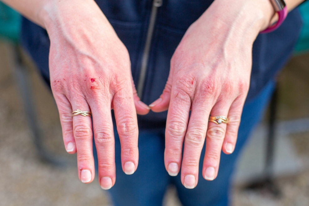
Among the most conspicuous signs of basal cell carcinoma is the form of lumps diagnosed on the skin. These lumps are generally tiny and solid and can look as if they have a pearly feel. At first, it might not be seen to be small. Thus, it might be considered a benign skin growth formation in the chest and arms of a woman. Yet, they are to stay or progress slowly over time, which makes it clear that there is a specific problem.
The lumps can be like looking through tissue paper. They may show small blood vessels under the skin, a sign that basal cell carcinoma is present. Lumps are also pretty much transparent but these lumps are not found in any other skin lesions. They usually occur in the face and sunglasses/wearing a hat might help or on the neck and/or the shoulders. These areas have the most exposure to UV radiation because they are the most vulnerable to UV rays.
That will finally result in the lumps turning ulcerated or completely scabbing over. That might be caused by an insignificant injury unnoticed to one’s consciousness. A small scratch can be easily mistaken for a small injury or a scab. The most distinguishable feature of these lumps is that they do not heal properly. They quickly break when damaged or bumped and sometimes even begin to bleed.
2.
Red-flat-colored skin surfaces are distinct among the symptoms of basal cell carcinoma in the early stage. Some of these areas can be flat. Others may be raised and of irregular shape. They look rough and cuspidate like a person who experienced allergen effects and got eczema. Conversely, the patches formed through the above-mentioned treatment may be similar. However, they typically last for weeks or months and are not relieved by lotion or moisturizer.
Initially, these patches may be pink or red and may progressively become more vivid red ones. Those endowed by heaven and at the peak of health are still energetic and young. In addition to a few itchings and irritations, those who suffer from these patches may also feel milder to moderate pain in those areas. However, the pain is not as high as in most other skin diseases. A continuous and rundown path is the precursors of basal cell carcinoma, and these might be something more threatening.
Hence, they thicken and become more raised and turn into roughness or scaliness in the redish ways. The variation might not be so apparent. Therefore, in the early stages of misdiagnosis, it might happen if one takes the skin patches as a symptom of dry skin or mild dermatitis.
3.
Producing a scar-like basal cell carcinoma (BCC) appears to be one of the lesser-known symptoms. It can be challenging to recognize this sign as it looks like a flat, smooth piece of skin or scar not associated with an injury. The occurrence of this stuff as either morphea form or sclerosing basal cell carcinoma is a regular case. Consequently, the early stage of this disease would not be detected, which, in turn, will lead to a delayed diagnosis. This type of growth is often confused with a benign scar, and hence, people go to the doctor for a check-up quite late.
The most typical color of these areas, which look like scars, is white, yellow, or skin-colored. They are smooth, waxy, or shiny on their surfaces. The patches themselves do not resemble normal scars that are a direct result of injury or surgery. They form themselves over time and start spreading out.
Even though these wart-like scars have no symptoms, sometimes there is an uncomfortable or tight feeling over them. Basically, these clues, along with the stranger nature of such growths, motivate people to ask for medical consultation from a professional if such an area is noticed with no clear cause.
4.
Next, another indication of the existence of basal cell carcinoma is the appearance of a pink growth. Raised platforms highlighted by flame ulceration could be common in pink growth, which wicked around and appears to be unifoliate at some points, among other cyclical patterns, and often shaped like a finger whose low center is like a funnel. The aforementioned growth will be very noticeable. Many times, the growth is called a rodent ulcer because it has an unattractive look and causes a slow rate of growth.
The pink growth is described as a lace pattern made up of vessels so small that they are hardly visible, creating the image of a delicately patterned quartzoid in a virtual world. The lesion is of pink color, and besides being vascular, it is an identifiable or indicative basal cell carcinoma that comes with it. The pink growth is soft when felt, and this gives it a texture that differentiates it from masses that have not been previously removed.
With the progress of the pink growth, it may reach the stage of ulcerating or crust in the middle; thus, it may also bleed or produce an artificial scab off and on. Almost invariably, terms would be mistaken to view these changes as sores or wounds that refuse to be healed and would, therefore, not give a correct diagnosis. However, they are very characteristic of basal cell carcinoma as they never seem to go away, although they sometimes become inflamed or produce a scab on their surface.
5.
The appearance of shiny bumps or nodules in the skin is the most common sign of basal cell carcinoma, also known as skin cancer. They are the most common type of skin cancer and the least severe. They cannot be easily cured without the right treatment. In fact, after many years of love and care, they may remain the same. They can also refuse to budge at all. They can look as if the only option is the amputation of the infected tissues.
The sores on the sides may not only get thick but also grow inwards so that the center appears sunk in. The characteristic that makes basal cell carcinoma more than just another kind of skin cancer is the growth of sores that never heal or the spread of an injury. The sore might be tender and may display mild pain or discomfort, especially as it increases irritation or friction caused by skin-to-skin contact in areas such as the hands or face.
With time, the open sore, which is one of the most dangerous results of abnormal cell growth and differentiation, might not only surpass the initial sole but also represent the tendon, muscle, and bone in the deeper layers that the initial wound chose to invade. In these cases, the doctor must do everything he can to eradicate the infection, even if it means that the patient receives medical attention. If the sore is left untreated, it can result in severe complications. These involve the infiltration of deeper structures such as muscles and bones.
6.
A shiny bump or nodule is usually the first sign of basal cell carcinoma in the form of a round, shiny growth on the skin. These growths are characterized by a pearly white or pinkish hue. Darker shades like brown or black type and the presence of a dull and rough texture can also be observed at different stages of their development based on skin color. Mentioning these features, doctors are not concerned about possible mistakes or faults if those lesions are not biopsied. The change in color and longevity sets it up as the most predominant nodular lesion on sun-exposed body areas. The papular types only follow.
Moreover, the most remarkable thing about them is that they look as if you were looking through the skin. You could see the blood vessels inside. That is not the case with nodules. They are smaller than skin bumps and growths, and they do not grow slowly like cancerous tissue. They occur primarily in sun-exposed areas like the face, ears, or scalp.
The new comes when the shiny bumps look like ulcers or may have a depressed central portion as happens with the other basal cell carcinomas as well. When bleeding takes place persistently, the patient should see a specialist.
7.
In the early stages of basal cell carcinoma, a part of the skin that is irritating and does not seem to heal properly after conventional treatments can be considered fatigue beside an internal organ. They could occur in the form of red, dry, or watery patches on the skin that would last for weeks or months and still not be completely gone. Most of the time, they are probably wrong as they are usually diagnosed as psoriasis or contact dermatitis, to which ointments and anti-inflammatory medications are prescribed as treatment instead of the correct medication for the diagnosis.
The mild irritation can either be a constant one or appear sporadically. Some patients may have itching, burning, or pain only in some specific areas. These red areas – very different from other infections seen on the skin – are less responsive to ointments. They also would not heal when anti-inflammatory medications are used. In addition to this, the way they look also emphasizes the fact that they are other types of skin infections.
Along with the development of irritation, the area may also be accompanied by other signs. These are a higher sheen and gloss, more blood vessels visible, and the height of the edge. These features continue to magnify the mandatory monitoring of persistent cutaneous irritation and the subsequent referral to a dedicated physician who is an expert in the field of skin diseases and their treatments and specifically has experience in this area.
8.
Basal cell carcinoma is a skin tumor that develops on the outermost layer called the epidermis. It is the praise of some small red blood vessels because of the presence of basal cells in the layers beneath the epidermis. Telangiectasia is a vessel disorder of the skin that originates from a vasculature malfunction of a body part situated close to the diseased one. Therefore, it can be considered a sign of basal cell carcinoma. These feathery capillaries can occur in a patch and form a peculiar web pattern. The vascularity of the neoplastic lesion is higher than that of the normal skin. Small vessels are easily noticeable, and they look much less than in normal skin, although there is no great difference.
These redness (the red vessels) become more apparent with cutaneous nodules or growths that are shiny and translucent. On the other hand, the color of the vessels becomes light to dark. The tissue content varies, translating from blue or violet to red on the skin surface or perhaps into deeper structures. It is also thin and has a vulnerable look which is right and essential for the dermatologists in the diagnosis process.
More and more of these small blood vessels are likely to become visible. Bleeding from the lesion is very likely to occur as well. The adjacent acute blood vessels may also display minor icterus due to the irritation from localized trauma of the microcirculation. This will lead to the wound bleeding or forming a scar around it due to the trauma. Though it may be small bleeding, an immediate medical consult and therapy are needed for the bleeding and the ruptured vessels. It is because this could be a sign of a more serious condition.
9.
Ulceration is essentially a learning curve of basic knowledge and more complex than grounds of scattering. It is the reason that beyond the normal expansion and the penetrating through the deeper layers of the tissue. It is manifested by the continuous growth of the carcinoma of the skin. Include no healing of central depression or erosion in the lesion. The beginning of the process is the central depression in the wound. Later, the wound could become an ulcer.
The ulcerated base wrinkles and the outline create a well-visible, elevated, and slit-like rim, which is not typical of other skin areas that are flat and practically regularly structured. A local anesthetic agent can help in taking an ulcer off. Alongside this, it can be not comforting to see raised borders present. Walking on soles can ooze or be crusty due to friction or small vessel bleeding.
Indeed, ulceration is the one that is usually associated with unpleasant sensations and a variety of complaints that range from the mildest to the most severe pain, the depth of the ulcer in this case is the deciding factor as to the degree of discomfort. Seldom May skin during basal cell carcinoma development grow less rapidly and the first period may be like this one. However, ulceration marks the chronic phase.
10.The first or subclinical sign of basal cell carcinoma is a small discoloring of the skin (lighter pigmentation). That can be a very early symptom of basal cell carcinoma in the morphoeaform subtype. All the hairless, smooth, and flat areas are the places for symptoms, such as whitened skin areas, which are created after the appearance of the first signs. The color changes might be occurring. However, it is a fact that most often, you can’t see the skin’s mild changes before it is through visualization techniques or tricking ontological devices of the dermatologist.
Whereas a bright and shiny feel identifies abnormal areas like chloasma, the linear defect of the pigmented layer with the help of a slit lamp, for instance, is less obvious. Meanwhile, papular formations and other skin changes are distinguished by smooth and shiny surfaces that are free of pigment. This image might accept the fact that the space between the formation and the skin remains almost imperceptible, causing some areas to be fused.
The regions or areas of the skin can actually be the early signs of basal cell carcinoma, while otherwise, the doctor may suspect other also non-malignant problems such as little blood vessels or some local irritation. The top layer of the skin can become so light that it seems almost like white tissue paper. If there is no pain but the area increases in size and persists, then submit to the medical consultation to make sure you are not ignoring any health threats that could be related to it.