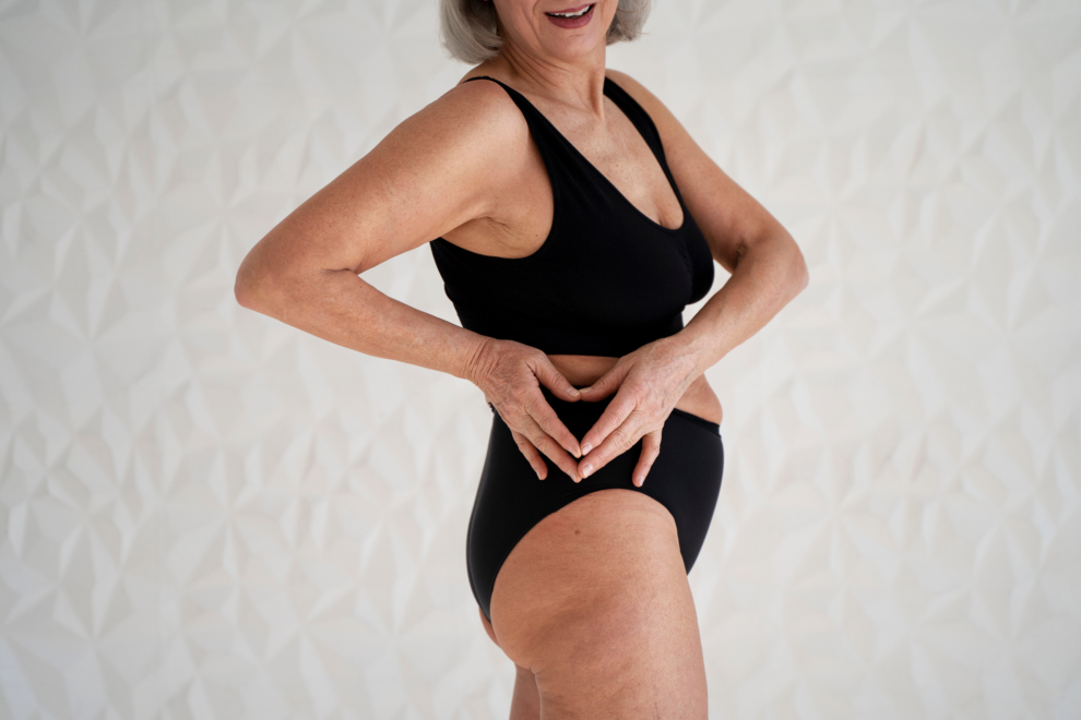
The most common cause of lateral hip pain is trochanteric syndrome (formerly known as greater trochanteric bursitis). It may be associated with bursitis, caused by tendinopathy or a tear/rupture of the gluteus medius tendon. Iliotibial band friction syndrome and external snapping hip syndrome can contribute to the severity of trochanteric syndrome.
The trochanteric syndrome presents with lateral hip pain that is worsened by walking or other physical activity, prolonged sitting, and sleeping on the affected hip. It most often affects women aged 40–60.
Usually, there is no identifiable cause of the injury. The patient may have a Trendelenburg gait or have a positive Trendelenburg or external derotation test against resistance.
2.
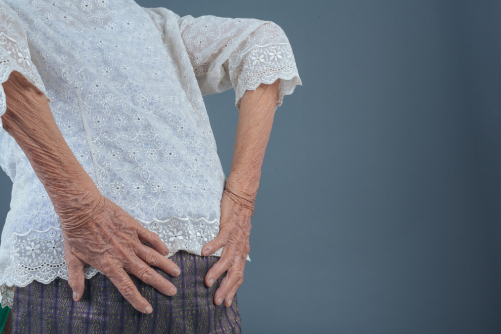
A fracture of the femoral neck is a common problem in the elderly population, which manifests itself with severe pain in the groin, intensifying when trying to walk, and visible shortening of the limb. In most cases, the recommended procedure is surgical treatment, the aim of which is to stabilize the fracture and enable movement without hip pain.
Most such fractures concern elderly people. Seniors often suffer from osteoporosis and their bones are not as strong as those of young people. Weakened bone tissue causes a fracture even with so-called low-energy trauma, i.e., with injuries that seem harmless, e.g., falling from one’s height (falling over). In younger people, much greater force is necessary for the formation of this type of fracture, such as, for example, a fall from a great height, or a road accident. Such a fracture rarely occurs without trauma, in the mechanism of the so-called fatigue fracture, i.e., when overloading of a given area is repeated, as in athletes who train frequently and more intense. In addition to older age, other risk factors for hip fracture include:
A hip fracture is characterized by severe groin pain and limited hip mobility. A patient with a fracture cannot stand on the hurt leg, and any attempts to move it intensify the symptoms. After a fracture, there may be a visible shortening of the injured limb relative to the healthy side.
3.
The bursa is a sac with fluid that facilitates muscle movement over bones and lubricates joint cartilage. There are 150 bursae in our musculoskeletal system. The hip joint has the largest of them – the trochanteric bursa, located between its posterolateral surface and the gluteus maximus muscle (the outer area of the hip). This bursa supports the work of the quadriceps femoris muscle, which flexes the hip.
Bursitis means inflammation, swelling, fluid accumulation, and the resulting complex movements of muscles and tendons.
In the initial phase of inflammation, pain is usually not felt but the lack of diagnosis and continued physical activity leads to an imbalance between the muscles of the hip joint, and then hip pain appears.
4.
A hip fracture is a serious orthopedic injury that affects seniors and people with osteoporosis. The hip is one of the largest and most mobile joints in the human body, and a fracture can occur in any part of it, although it is usually the neck or trochanter of the femur. Each case requires appropriate physiotherapy to return to full fitness.
There are also several types of hip fractures depending on the location of the fracture gap. The most commonly diagnosed fracture is the femoral neck fracture, less frequently a fracture of the acetabulum, a pertrochanteric fracture of the femur, or a fracture of the sub-capital of the femur. If the snapping in the hip area is sporadic, painless, and does not impede normal functioning – it does not require medical consultation. In the situation when the snapping is persistent, causes discomfort, is painful, or interferes with the patient’s professional or everyday activities – then consultation at an orthopedic clinic is necessary.
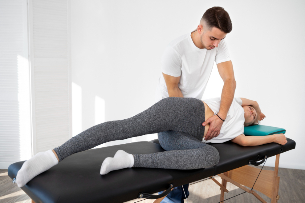
If the problem is not correct posture, contractures, and muscle imbalance (imbalance in the tension of individual muscle groups), physiotherapy brings the best results. Correction of posture defects, stretching, and elasticity of contracted muscles through appropriately selected exercises and manual therapy gives a chance to effectively solve the problem. In exceptional cases, when physiotherapy does not bring the effect and the problem significantly impedes functioning, surgical treatment can be considered, usually using minimally invasive methods. If the cause of the snapping hip is intra-articular pathology, treatment is much more difficult, and surgical intervention is much more often essential. Arthroscopy of the joint may be necessary to repair the labrum, remove a loose body, etc. More extensive procedures may be indicated less often.
5.
Osteoarthritis of the hip joint is primarily associated with the wear and tear of cartilage and the formation of abnormal bone growths known as osteophytes. These changes typically occur as part of the aging process and are referred to as primary degenerative hip joint diseases. Osteoarthritis can also develop as a result of childhood joint diseases (such as dysplasia), injuries, and certain systemic conditions (like rheumatoid arthritis). It is referred to as a secondary degenerative disease of the hip joint, which occurs less frequently.
The most common symptom is pain in the area of the joint (i.e., hip pain), which can also radiate to the knee joint. It can prevent normal sleep. The symptoms usually start mildly and become more severe over time. During everyday activities, joint stiffness and a limitation of its range of motion are often felt. In addition, the patient may experience an unpleasant sensation of creaking in the joint, which is caused by the destruction of the normal cartilage surface and may even result in a complete limitation of joint mobility.
All of these symptoms lead to a decrease in overall efficiency, which contributes to poorer functioning both in the professional sphere and in everyday duties. The course of coxarthrosis is usually progressive. If the above symptoms appear, a planned visit with an orthopedic doctor is recommended. It should not be postponed because the natural development of coxarthrosis leads to advanced degenerative changes and a significant decrease in the quality of life.
6.
The iliotibial band is a tape-like structure that runs along the outer side of the thigh. Its role is part of the fascia, which helps maintain the proper positioning of muscles. At its lower end, located on the lateral side of the knee, the iliotibial band moves freely over the bony prominence known as the lateral epicondyle of the femur during activities such as running or walking.
Iliotibial band syndrome (ITBS) occurs when the natural movement of the band becomes painful and disrupts normal functioning. Iliotibial band syndrome most often affects runners, cyclists, and other athletes in disciplines that require squatting and repeated knee bending.
The anatomical structure of the lower limbs may predispose to the syndrome. However, the most significant factor is general motor preparation and training habits. Poor flexibility of the lower limb muscles, imbalance between different muscle groups, and poor pelvic stabilization are risk factors related to preparing the body for workout Training errors that can lead to the onset of this syndrome include overtraining, insufficient recovery, excessive uphill or downhill running, running on uneven surfaces, and running on inclined terrain. Additionally, various inflammatory processes in the body may also contribute to the development of this syndrome.
The primary symptom of iliotibial band syndrome is pain on the outer side of the knee, which may radiate toward the hip. This pain often starts during or after exercise and can become chronic, sometimes constant. It typically does not occur at rest but may arise when bending the knees—such as when walking up stairs. Some individuals also report pain below the knee, which can radiate to the calf or higher up toward the thigh. Other symptoms may include swelling in the lateral knee area, a crunching or jumping sensation in that region, tingling, and a burning sensation.
7.
Hip and pelvic joint overload is common among athletes, physically active individuals, and manual laborers. Because these structures are closely located and interconnected, a problem in one can lead to dysfunction in the other. Static and dynamic overload of the musculoskeletal system are frequent causes of pain in the lower limb joints.
Excessive tension of the soft structures of the pelvis and hip joint can lead to overload not only them but also joints or bones. Usually, the cause of such discomfort is located in a distant area, e.g., the abdominal cavity or spine. Often, the joint capsule of the hip joint is subject to increased tension, which is most activated in the standing position. It is thick and strong and attaches to the pelvic bone, and one of its tasks is to relieve the muscles of the hip joint.
It seems obvious, but sometimes we can forget about it. Hip pain should not be ignored and remember that it is much easier to get rid of if we do not worsen the injury. Of course, rest does not mean have to give up physical activity and lie in bed. On the contrary, the hip cannot “stall”, because due to a complete lack of movement, it can be stiff and hurt even more. However, you should give up exercises that strongly engage the hips, such as running, and instead go to the swimming pool. Proper nutrition is also essential. Overweight and obese people should reduce their body weight, as it affects the overload of the hip joint.
8.
Hip dysplasia is characterized by the inadequate development of the bones and structures that form the hip joint, particularly the acetabulum (the “socket”) on the hip bone that connects with the femur. This condition causes the femur, as the muscle strength and tension increase, to be permanently pushed out of the acetabulum outside the joint. It leads to a joint dislocation, which prevents the child from walking properly.
If left untreated or treated too late, a child with hip dysplasia may develop a limp, often resembling a duck walk. The condition is more commonly unilateral (affecting one hip) but can also occur in both hips. Hip dysplasia is more prevalent in girls than boys, and the risk is higher for children whose siblings or parents have had the condition or for those who were born in a breech position.
When detected early, hip dysplasia can be effectively treated using non-surgical methods. However, if a hip dislocation occurs, surgical intervention may be necessary, followed by long-term immobilization in a cast. Treatment for these hip conditions should be managed by a pediatric orthopedist.
9.
Hip joint inflammation is more common in children aged 3 to 10 years, especially in boys. This condition often arises as a result of bacterial or viral infections. It can occur as a post-infectious complication following upper respiratory tract illnesses, such as tonsillitis or the flu—both are frequent in childhood.
Even seemingly minor infections should not be overlooked, as untreated infections can lead to severe inflammation in the hip joint. In addition to transient hip joint inflammation, there are two other types: bacterial (infectious) hip joint inflammation and viral hip joint inflammation.
Clinical symptoms of transient hip joint inflammation include:
Symptoms of infectious (bacterial) inflammation of the hip joint include:
Characteristic symptoms of viral inflammation of the hip joint include:
Bones can have tumors of various types – malignant or benign. Bone tumors usually originate from cartilage, bone, or fibrous tissue. Non-cancerous (i.e., benign) bone tumors are much more common than cancerous tumors. They can occur at any age, including in children. Benign bone tumors do not cause metastases, do not pose a health risk, and their treatment usually consists of removing the lesion. Some benign bone tumors do not require treatment, it depends on the location of the change and the symptoms associated with it.
A malignant bone tumor usually causes pain and swelling in the area of the change. The pain may not be constant but may come and go, and usually intensifies over time – over weeks or months. The pain may intensify at night and during physical exercise. In some people, the tumor may cause visible thickening of the limb or can be felt under the skin. Bone sarcomas may be accompanied by limb dysfunction, i.e., limited mobility of the nearest joint and its reflexive sparing by the patient, which may manifest itself, for example, by limping. In advanced sarcomas, the bone may break at the tumor site with a minor injury that would not result in a fracture of the healthy bone. It is important to remember that similar symptoms can be caused by other diseases.
Treatment depends on the type of bone cancer. Treatment methods include: